
The case file: why these simulators stall
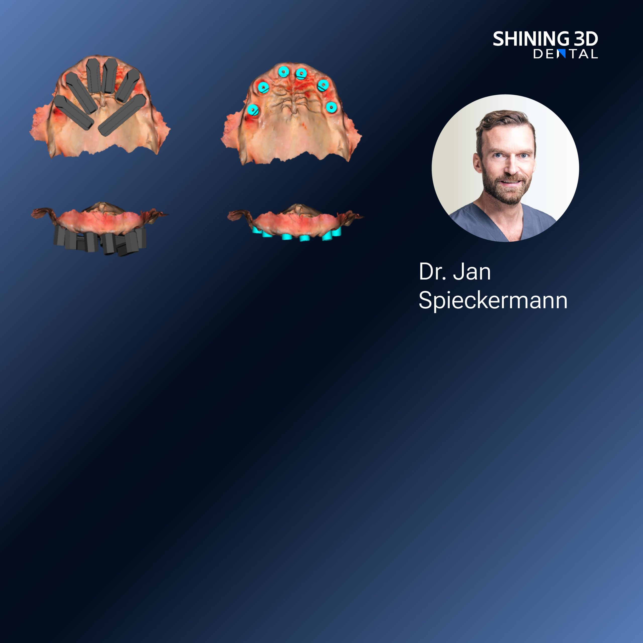
There’s a pattern to stalled straight-teeth simulations, and it begins at the scanner. A mismatched scan, a jitter in tracking, a missing bite—any of these leave a trail. Teams using a modern Photogrammetry 3D scanner know the capture phase can be precise, but only when every step lines up. Treat the first scans like fingerprints: if they’re smudged, nothing that follows will be reliable.
Problem diagnosis: three recurring breakdowns
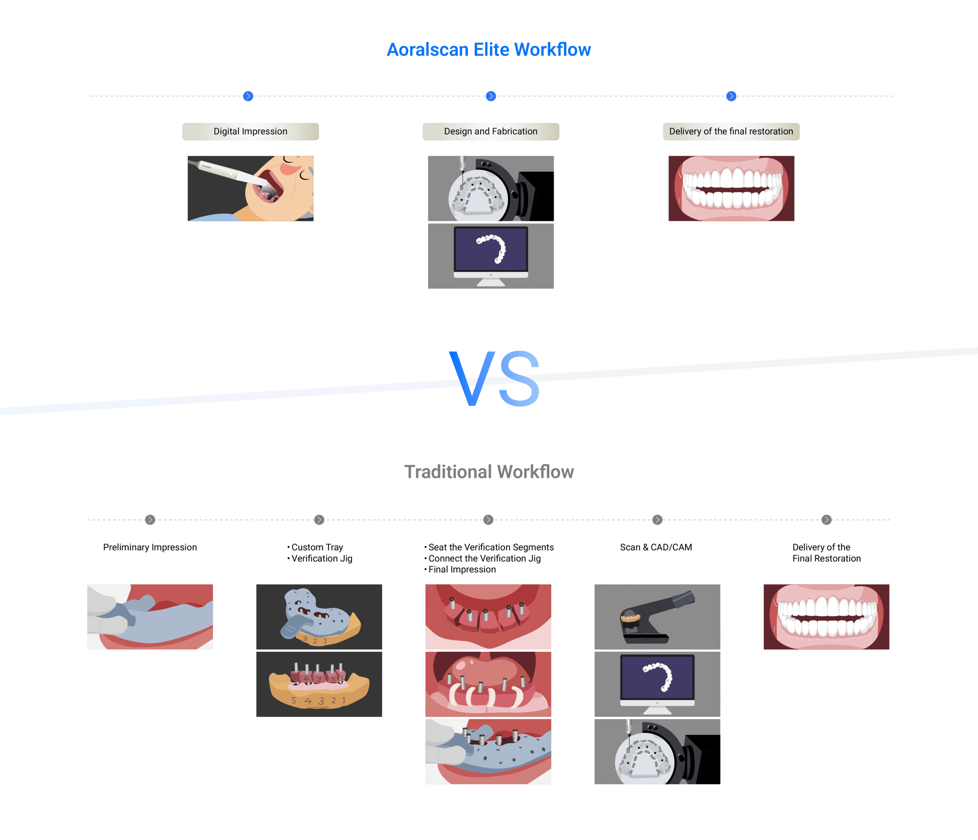
1) Input inconsistency: variable lighting, patient movement, or tooth moisture create noise. 2) Integration gaps: file formats and metadata drop out between capture, CAD and aligners. 3) Validation blind spots: teams accept a visual fit without objective metrics. These three issues explain most failed cases. Find which one shows first and the rest fall into place.
Collecting the evidence: what to measure
Measure capture repeatability, model-to-model deviation, and time-to-clinical-decision. Track how often a scan requires re-capture and document why. My experience with multi-site clinics and lab partners shows that tracking these three metrics reduces rework quickly. These are practical measurements: millimeters, minutes, and re-capture counts—not vague satisfaction scores.
Real-world anchor that matters
At the IDS show in Cologne I observed intraoral photogrammetry workflows demonstrated on live models; those demos highlighted the same variables teams face every day. That day confirmed what field testing suggests about intraoral photogrammetry / IPG: it excels where capture protocol is strict and fails where teams improvise.
Remedies: step-by-step fixes that work
Start by standardizing capture: fixed lighting, explicit moisture control, and a short scripted patient instruction set. Adopt a single primary file format and require metadata (bite stage, scan orientation). Add a two-stage QA: an automated geometric check (mesh overlap < threshold) followed by a blinded visual pass by a trained technician. Train staff in short, focused sessions and log every re-capture with a reason code. These concrete steps turn sporadic success into predictable outcomes.

Alternatives and their trade-offs
Photogrammetry provides high-accuracy full-arch captures with minimal intraoral bulk; traditional intraoral scanners are faster for quadrant work and more forgiving for movement. CBCT gives hard-tissue context but is overkill for routine alignment planning and adds radiation. Choose based on the case type: full-arch aligner plans favor photogrammetry when capture discipline is present; small posterior adjustments often don’t need it.
Common pitfalls teams repeat
Rushing capture, skipping metadata, and assuming visual acceptability equals clinical acceptability. Also, ignoring version control between CAD revisions and manufacturing files creates silent divergence—two teams think they’re working from the same model when they’re not.
Deployment checklist
– Define capture protocol and lock it in a written SOP. – Assign capture ownership per appointment. – Require metadata with every file. – Run a validation case weekly with measurable thresholds. – Keep a single source-of-truth file repository with versioning. – Train and retrain on edge cases (severe crowding, restorations).
Closing the case
Teams that treat simulator workflows like an investigation—collecting hard evidence, testing hypotheses, and correcting the primary error—stop chasing symptoms and start delivering predictable results. Where protocols are clear and measurement is constant, solutions form quickly, and that practical rigor is what differentiates routine wins from costly rework, as seen in field trials and supplier demos from trusted vendors such as SHINING 3D DENTAL.